Glioma is one of the most common types of cancer of primary brain tumors. It develops in the brain and spinal cord. They start in glial cells that surround nerve cells and help them function. Depending on the type of glial cell it affects, the characteristics and type of tumor will vary.
In our post on monoclonal antibodies for cancer, we told you about some of the mechanisms and examples that these antibodies use to try to fight this type of disease.
In the case of glioma, many studies have shown that the blood-brain barrier allows the entry of monoclonal antibodies, which is why they are considered a good starting point for the treatment of intracranial malignant cells.
For the investigation of the glioma, antibodies that are directed, for example, against different growth factors are used, which we will discuss later. The study of these biomarkers plays a fundamental role in the development of the disease.
3 MONOCLONAL ANTIBODIES FOR THE STUDY AND TREATMENT OF THE GLIOMA:
Bevacizumab
As we mentioned in other posts, this monoclonal antibody attacks the vascular endothelial growth factor (VEGF) whose main objective is to prevent angiogenesis, the generation of blood vessels by the tumor to obtain nutrients and promote its growth.
In this case, Bevacizumab, also called Avastin, is mainly used for glioblastomas. It can be used alone or combined with chemotherapy by intravenous infusion.
Everolimus
This antibody promotes reduction of tumor size or growth rate. It binds to a protein cell, mTOR, which promotes cell growth and division.
This type of antibody is administered in pill form, in most cases.
Nimotuzumab
In this case, nimotuzumab is also directed against a growth factor receptor, in this case the endothelium (EGFR), inhibiting its growth. Furthermore, it promotes apoptosis, limits tumor neo-angiogenesis and has radiosensitizing action. This is why this antibody is often used in combination with radiation therapy.
If your research project is related to this disease, at Abyntek we have a multitude of monoclonal and polyclonal antibodies for the study of glioma against biomarkers of this type of tumor.
The predominance of high abundance proteins in biological samples, often complicates the work to analyze and detect low abundance proteins known as LAPs (low abundance proteins). In the case of plasma, for example, 99% of its total protein content is made up of only 22 major proteins, while the remaining 1% corresponds to hundreds of proteins that are expressed in low amounts.
Standard methods can be used to detect low abundance proteins in a biological sample, but these require previous steps of optimization and signal amplification.
In this entry we summarize the main methods for detecting low abundance proteins in biological samples.
METHODS TO DETECT LOW ABUNDANCE PROTEINS
Before proceeding to the analysis of the low abundance proteins, it is necessary to carry out a process of enrichment of the sample by depleting the highly abundant proteins in it, thus increasing the specific signal of the low abundance proteins, facilitating their detection.
There are several methods to enrich these samples, including:
Collagenase treatment (can double the detection signal of low abundance proteins)
Isopropanol extraction with polyacrylamide gel electrophoresis
Polyethylene glycol separation and immunoaffinity-based depletion (this method uses IgY antibodies and can increase sensitivity up to 3 times)
Protamine sulfate precipitation combined with Western Blot (simple and low cost method to deplete abundant proteins)
Magnetic nanoparticles
Capture reagent-based immunodepletion methods
Immunoaffinity partition
Heparin chromatography (can increase detection signal by more than 20%)
Once the sample has been enriched, the detection method that will be used to detect low abundance proteins must be optimized:
MASS SPECTROMETRY
Signal augmentation mass spectrometry is one of the most widely used methods to detect low abundance proteins in various types of samples, especially in serum, adipose tissue, bone and tendons.
This analysis can be improved using affinity capture reagents against the protein of interest, since this allows to increase its concentration.
WESTERN BLOT
The properly optimized western blot is a very effective method to detect low abundance proteins. The optimization of this technique goes through:
Selection of the appropriate lysis buffer
Protein precipitation
Optimization of gel electrophoresis
Optimization of the concentration of the primary antibody
Optimization of signal detection
ELISA
Although standard ELISA assays are not sensitive enough to detect low abundance proteins, combining this technique with a proximity ligation assay (PLA), it is possible to significantly increase the specificity when detecting the protein of interest simultaneously by means of numerous probes.
This method has been used for the detection of biomarkers of neurodegenerative and inflammatory diseases.
NUCLEAR MAGNETIC RESONANCE
Another of the most effective methods to detect low abundance proteins is high resolution nuclear magnetic resonance, which offers high sensitivity and selectivity without the need for a previous isolation procedure.
This method has been used for the analysis of Alzheimer-related biomarkers such as the β-amyloid protein.
Fifty one sufferers with abnormalities of horizontal gaze had been studied with magnetic imaging of the mind (MRI) and eye motion recordings to determine the loci of lesions chargeable for remoted abducens palsy, conjugate gaze palsy and differing kinds of internuclear ophthalmoplegias.
The lesions chargeable for a halficular dysfunction had been recognized by overlapping enlarged drawings of the person scans at comparable brain-stem ranges and figuring out the areas the place the irregular MRI indicators intersected.
A statistical process was devised to exclude the chance that the areas of overlap occurred by likelihood. In this paper, the findings within the group of sufferers with VI nerve palsy are reported because the location of their lesions may very well be predicted from identified anatomy, so validating the process.
The outcomes had been independently obtained with the overlapping approach and the statistical process and confirmed that the lesions had been positioned in a area comparable to the posterior half of the abducens fasciculus.
This confirms that central lesions producing remoted lateral rectus weak point spare the abducens nuclei. The settlement between the procedures used and earlier medical and experimental outcomes recommend that the strategy we describe might be utilized to find the location of lesions on MRI scans in different teams of sufferers with extra complicated gaze issues.
Abnormalities of horizontal gaze. Clinical, oculographic and magnetic resonance imaging findings. I. Abducens palsy.
Thyrotropin-releasing hormone inhibits GH4 pituitary cell proliferation by blocking entry into S section.
TRH inhibits the proliferation of GH4 rat pituitary cells. We have characterised TRH inhibition of cell proliferation by 4 approaches: cell quantity, [3H]thymidine incorporation per tradition, bromodeoxyuridine (BrdUrd) incorporation per cell, and cell cycle distribution. TRH decreases GH4 cell quantity inside 18 h of remedy, and this inhibition is maintained for as much as 96 h. TRH inhibits [3H]thymidine incorporation into GH4 cell cultures as early as 12 h, and the inhibition of [3H] thymidine incorporation correlates, after a 6-h lag, with decreased GH4 cell quantity.
TRH inhibition of [3H]thymidine incorporation is focus dependent and saturable, with half-maximal inhibition (IC50) of 2 nM. TRH inhibition of [3H] thymidine incorporation is receptor quantity dependent as much as 160,000 websites/cell, suggesting no spare receptors for TRH on GH4C1 cells.
The exact motion of TRH on GH4 cell proliferation was examined by move cytometry of fluorescein isothiocyanate-anti-BrdUrd- and propidium iodide-DNA stained cells. TRH inhibits the quantity of cells that incorporate BrdUrd and not the quantity of BrdUrd included per cell.
Dual evaluation signifies that the decreased anti-BrdUrd staining is basically restricted to cells within the early S section. This motion of TRH is extended (better than 32 h) and ends in a parallel improve within the quantity of cells in G2-M and G1. These findings point out that TRH inhibits GH4 cell proliferation at the very least in half by inhibiting the quantity of cells coming into the S section.
Three-dimensional (3D) printing could also be an answer to shortages of gear and spareelements within the healthcare sector of low- and middle-income nations (LMICs). Polylactic acid (PLA) for 3D printing is broadly obtainable and biocompatible, however there’s a hole in data regarding its compatibility with chemical disinfectants.
In this examine, 3D-printed PLA tensile samples have been created with six completely different printer settings. Each of these six batches consisted of 5 units with 5 or 6 samples. The first set remained untreated, the others have been soaked in Cidex OPA or in a chlorine answer.
These have been utilized for seven consecutive days or in 25 brief cycles. All samples have been weighed earlier than and after remedy and subjected to a tensile check. Results confirmed {that a} third of the remedies led to a rise of the median weight with a most of 8.3%, nonetheless, the samples with one of the best floor high quality didn’t change.
The median energy enhance was 12.5% and the biggest lower was 8.8%. The median stiffness decreased 3.6% in a single set and elevated in three others as much as 13.6%. When 3D printing PLA medical instruments, floor porosity should be minimized to stop switch of disinfectants to folks.
The large variability of mechanical properties attributable to 3D printing itself and as a consequence of disinfection should be thought of when designing medical instruments by deciding on applicable printer settings.
If these circumstances are met, reusing 3D-printed PLA medical instruments appears protected from a mechanical level of view.
Protection impact of cerium oxide nanoparticles in opposition to radiation-induced acute lung accidents in rats.
Radiation remedy is one of the commonest instruments for treating most cancers. The goal is to ship sufficient doses of radiation to kill most cancers cells and probably the most difficult half throughout this process is to guard regular cells from radiation.
One technique is to make use of a radioprotector to spare regular tissues from ionizing radiation results. Researchers have pursued cerium oxide nanohalficles as a therapeutic agent, attributable to its numerous traits, which embrace antioxidant properties, making it a possible radioprotector.
One hundred rats have been divided into 5 teams of A) management group, intraperitoneal (IP) saline injection was executed twice every week; B) bi-weekly IP injection of 14.5 nM (0.00001 mg/kg) CNP for 2 weeks; C) a single entire thorax radiation dose of 18 Gy; D) a single entire thorax radiation dose of 18 Gy + bi-weekly injection of 14.5 nM CNP for 2 weeks after radiation; E) bi-weekly IP injection of 14.5 nM CNP for 2 weeks previous to radiation + a single entire thorax radiation dose of 18 Gy. Thirty days after irradiation, 7 rats from every group have been anesthetized and their lungs extracted for histopathological examination.
Statistical analyses revealed that CNP considerably decreased the incidence of tissue collapse and neutrophile aggregation in rats receiving CNP earlier than radiation as compared with the radiation group.
The outcomes recommended the chance of utilizing CNP as a future radioprotector attributable to its means to guard regular cells in opposition to radiation-induced injury.
Members of the household of NEK protein kinases (NIMA-related kinases) have been described to have essential roles in regulating completely different features of the cell cycle. NEK10 was reported to take half in the upkeep of the G2/M checkpoint after publicity to ultraviolet mild.
NEK1, NEK5, NEK2 and NEK4 proteins alternatively have been linked to mitochondrial features.HEK293T cells have been transfected with FLAG empty vector or FLAG-NEK10 and handled or not with Zeocin.
For proteomic evaluation, proteins co-precipitated with the FLAG constructs have been digested by trypsin, and then analyzed through LC-MS/MS. Proteomic information retrieved have been subsequent submitted to Integrated Interactome System evaluation and differentially expressed proteins have been attributed to Gene Ontology organic processes and assembled in protein networks by Cytoscape.
For purposeful, mobile and molecular analyses two steady Nek10 silenced HeLa cell clones have been established.
Here, we found the next attainable new NEK10 protein interactors, associated to mitochondrial features: SIRT3, ATAD3A, ATAD3B, and OAT. After zeocin therapy, the spectrum of mitochondrial interactors elevated by the proteins: FKBP4, TXN, PFDN2, ATAD3B, MRPL12, ATP5J, DUT, YWHAE, CS, SIRT3, HSPA9, PDHB, GLUD1, DDX3X, and APEX1.
We confirmed the interplay of NEK10 and GLUD1 by proximity ligation assay and confocal microscopy. Furthermore, we demonstrated that NEK10-depleted cells confirmed extra fragmented mitochondria in comparison with the management cells.
The knock down of NEK10 resulted additional in adjustments in mitochondrial reactive oxygen species (ROS) ranges, decreased citrate synthase exercise, and culminated in inhibition of mitochondrial respiration, affecting halficularly ATP-linked oxygen consumption price and spare capability.
NEK10 depletion additionally decreased the ratio of mtDNA amplification, probably because of DNA harm. However, the whole mtDNA content material elevated, suggesting that NEK10 could also be concerned in the management of mtDNA content material.Taken collectively these information place NEK10 as a novel regulatory participant in mitochondrial homeostasis and vitality metabolism.
NEK10 interactome and depletion reveal new roles in mitochondria.
Ex vivo limb perfusion for traumatic amputation in army medication.
Limb loss has a drastic influence on a affected person’s life. Severe trauma to the extremities is widespread in present army conflicts. Among different features, “life earlier than limb” harm management surgical procedure hinders instant replantation throughout the brief post-traumatic timeframe, which is proscribed in half by the ischemic time for profitable replantation.
Ex vivo limb perfusion is at present being researched in animal fashions and reveals promising outcomes for its software in human limb replantation and allotransplantation.
The present lack of replantation prospects in army operations with excessive charges of amputation might be addressed with the event of a transportable ex vivo limb perfusion machine, as there are a number of alternatives current with the introduction of this method on the horizon.
We hypothesize that ex vivo limb perfusion will allow overcoming the important ischemic time, present surgical alternatives similar to preparation of the stump and limb, permit for spare–half surgical procedure, allow rigorous antibiotic therapy of the limb, scale back ischemia-reperfusion accidents, allow a tissue perform evaluation earlier than replantation, and allow the event of huge limb transplant applications.
Data from in vivo research in porcine fashions are restricted by the comparatively brief perfusion time of 24 h. In the army setting, notably longer perfusion occasions must be realized. Therefore, future animal research should focus particularly on long-term perfusion, since this represents the army setting, contemplating the time for stabilization of the affected person till evacuation to a tertiary therapy middle.
The growth and medical introduction of ex vivo limb perfusion in the army setting may result in a drastic discount in the variety of limb amputations amongst service members.
Ex vivo limb perfusion allows replantation surgical procedure in Role four amenities and adjustments the medical setting from a extremely pressing, life-threatening state of affairs to a extremely methodical, well-prepared place to begin for optimum therapy of the wounded service member. With its introduction, the precept of “life earlier than limb” will change to “life earlier than limb earlier than elective replantation/allotransplantation after ex vivo limb perfusion”.
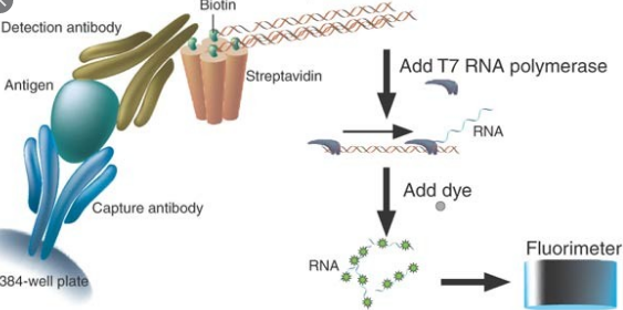
Healthcare providers, laboratory employees, and lab test developers must bear in mind that biotin, frequently seen in nutritional supplements, may interfere with certain strepravidin based laboratory tests and trigger erroneous test results that might go unnoticed.
Does the FDA Warns which Biotin-streptavidin may interfere with Lab Tests?
They recorded steptavidin-biotin interference in certain lab tests and offer recommendations for Labs, healthcare providers, laboratory personnel and laboratory test makers and programmers.
Considering that the 2017 safety communicating, some laboratory test developers are effective at mitigating the biotin interference of the assays, although some haven’t addressed it.
The FDA remains concerned about particular lab tests which haven’t addressed the chance of biotin-streptavidin interference. In a bid to improve transparency relating to this clinically significant hindrance, the FDA has made a decision to notify the general public about troponin assays who haven’t addressed the chance of biotin disturbance.
FDA recorded troponin In Vitro Diagnostic Devices that are subject to biotin interference but haven’t addressed this particular risk.
Similar problems arise from Macro b12 misinterpretation or Thyroid Function.
Reference:
Can Thyroid Function Measurements become atypical by Taking Biotin Supplements? (source Genprice Inc.)