Archives February 2022

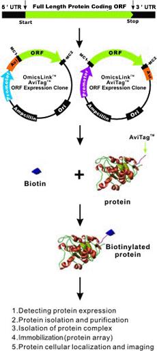
Cusabio Xenopus laevis Recombinant

Abstract

Details of the in vitro synthesis of double-stranded DNA complementary to purified Xenopus globin messenger RNA are presented, using a combination of reverse transcriptase, E. coli DNA polymerase 1 ‘A’ fragment and endonuclease S1. After selection of duplex DNA molecules that approximated the length of Xenopus laevis Recombinant globin messenger RNA by sedimentation of the DNA through neutral sucrose gradients, the 3′-OH ends of the synthetic globin gene sequences were extended. with short stretches of oligo dGMP using terminal transferase.

This material was integrated into linear pCR1 plasmid DNA extended with oligo dCMP and amplified by transfection of E. coli. Plasmids carrying globin sequences were identified by hybridization of 32P-labelled globin mRNA with total cellular DNA in situ, by hybridization of purified plasmids with globin cDNA in solution, by recombinant DNA analysis on polyacrylamide and agarose gels, and by heteroduplex mapping. The results show that extensive DNA copies of Xenopus globin mRNA have been integrated into recombinant plasmids.

Biological source: Xenopus sp.

Quality level: 100

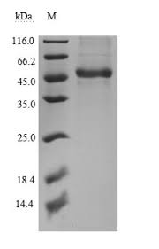
Molecular weight: molecular weight of 17 kDa

Manufacturer/trade name: Upstate®

Technique(s)

activity test: adequate

NCBI Accession Number: NM_002107.3

UniProt access no.: Q16695

Sent in: dry ice

Genetic information: Xenopus laevis…H3F3B(379757)

General description

Product Source: Produced in E. coli.

Quality

Routinely evaluated as a substrate for enzymatic reactions in vitro

Physical form

  • HPLC
  • Format: Purified

Storage and Stability: 2 years at -20°C

Other notes

For specific activity data, refer to the individual lot Certificate of Analysis for this enzyme.

Legal information

UPSTATE is a registered trademark of Merck KGaA, Darmstadt, Germany

Disclaimer

Unless otherwise stated in our catalogue or other company literature accompanying products, our products are intended for research purposes only and must not be used for any other purpose, including but not limited to unauthorized commercial uses. , in vitro diagnostic uses, ex vivo or in vivo therapeutic uses or any type of consumption or application to humans or animals.

Materials And Methods

  • Construction of the expression vector pUC18-PfHly III.

The codon-optimized PfHly III gene (GenScript, Piscataway, NJ; see Fig. S1 in Supplementary Material) was cloned into plasmid pET22b (Novagen-Merck Millipore). PfHly III was amplified from pET22b-PfHly III by PCR with a 5′ primer (5′-GGATCCCATCACCACCATCATCATGAATTCATGGAATTTTACAAAAAC-3′) and a 3′ primer (5′-TCTAGATCAGTGGTGGTTGGTGGTGGTG-3′) to generate BamHI and XbaI sites compatible with plasmid pUC18 (Agilent/Stratagene, Santa Clara, CA). The DNA insert was confirmed by sequencing.

  • Bacterial expression of recombinant PfHly III.

The ampicillin-resistant pUC18-PfHly III expression vector was transformed into competent Escherichia coli HB101 cells and cultured at an optical density at 600 nm (OD600) of 0.4, followed by protein induction at 37 °C of 16 h with 1 mM isopropyl-β. -d-thiogalactoside. The bacterial pellet was sonicated in 2 ml phosphate-buffered saline (PBS), followed by microcentrifugation at 12,000 × g at 4 °C.

recPfHly III was purified from the natively soluble supernatant by addition of preloaded Ni2+ nitrilotriacetic acid (NTA) resin and incubated overnight at 4 °C with gentle mixing by end-to-end rotation, followed by centrifugal washes with PBS containing 5 mM imidazole and finally elution with 100 mM EDTA. Elutions were dialyzed overnight at 4°C against PBS (pH 7.5) using Slide-A-Lyzer (Thermo Scientific) to remove EDTA and imidazole. Plasmid pUC18 alone was transformed, induced and purified under the same conditions and used as a negative control.

  • Western blot analysis of recPfHly III.

Soluble recPfHly III was separated by SDS-PAGE and transferred to nitrocellulose membrane (Bio-Rad). The membrane was blocked with Qiagen’s blocking buffer at 37 °C for 1 h, washed 3 times with PBS (0.05% Tween 20), and then incubated with a 1:1000 dilution of the anti-His-peroxidase conjugate. horseradish (HRP) on lock. buffer at 4°C overnight. The protein was visualized with enhanced chemiluminescence.

  • Hemolytic activity assay.

Human erythrocytes were washed with PBS (pH 7.5) three times and adjusted to a final concentration of 1% (vol/vol). The erythrocyte suspension (0.1 ml) was incubated with 0.1 ml recPfHly III diluted in PBS. The mixture was incubated for a total of 60 min at 37°C. The reaction mixtures were then centrifuged at 1,000 × g for 5 min.

Haemoglobin released from recPfHly III-induced hemolysis was monitored by the absorbance of the supernatant at 550 nm. One hemolytic unit (HU) was defined as the dose of recPfHly III that caused 50% hemolysis. One hundred per cent hemolysis was the amount of haemoglobin released after treating human erythrocytes (1%) with 0.1% Triton X-100. The recPfHly III hemolytic activity assay was studied at the indicated temperatures.

  • Osmotic protection experiments.

For osmotic protection experiments, 0.1 ml of recPfHly III (2 HU) was incubated with 0.1 ml of human erythrocytes (1% final concentration) suspended in PBS (pH 7.5) containing an osmotic protector at a final concentration of 30 mM (17). Incubation was at 37°C for 60 min and the mixture was immediately subjected to hemolytic activity assay. Osmotic protectants included glucose, polyethene glycol 600 (PEG 600), PEG 1500, PEG 2000, PEG 3350, PEG 4600, PEG 6000, and PEG 8000 (Sigma). The control used recPfHly III without any osmotic protector.

Cusabio N-terminal 6xHis-Avi-tagged Recombinant

Description

Recombinant human CD55 spanning amino acids 35-353. This construct contains a C-terminal Avi-Tag followed by a C-terminal His tag (6xHis). The recombinant protein was enzymatically biotinylated using N-terminal 6xHis-Avi-tagged Recombinant and affinity purified.

Construct: CD55 (35-353-Avi-His)-(biotin)

Species: Human

Host species: HEK293

MW: 38kDa

Glycosylation: This protein runs at a higher MW by SDS-PAGE due to glycosylation.

Genbank: #NM_000574

Tag(s): C-terminal Avi-His-Tag

Label: This protein is enzymatically biotinylated using Avi-Tag™ technology. Biotinylation is confirmed to be ≥90%.

Amino acids: 35-353

UniProt: #P08174

Storage stability: At least 6 months at -80°C.

Synonym(s): Molecule CD55, CR, CROM, Cromer blood group, DAFF, 100943-1, 100943-2

Purity: ≥90%

Formulation: 8 mM phosphate pH 7.4, 110 mM NaCl, 2.2 mM KCl, 20% glycerol.

Warnings: Avoid freeze/thaw cycles.

Scientific Category: Immunotherapy

Regulatory Status: For Research Use Only

Technology

AviTag™ technology is based on the biotinylation of AviTag™ by biotin ligase in vitro or in vivo and the specific and reversible binding of avidin or streptavidin to biotin to immobilize, purify and visualize proteins.

Combined with OmicsLink™ expression-ready clones

GeneCopoeia offers AviTag™ technology in a wide range of expression vectors. For example, it has been combined with the IRES (internal ribosome entry site) element and SUMO, 6xHIS, and a variety of other tags.  The AviTag with promoters driven by T7 or CMV is available for more than 20,000 human and 15,000 mouse genes.

Specific

The biotinylation of the recombinant protein carried by the AviTag is highly specific. In the presence of biotin and ATP, biotin ligase catalyzes the amide bond between biotin and the peptide-specific lysine 15-aa AviTag. Biotinylated proteins in nature are extremely rare, which makes the chances of cross-reactions, especially compared to antibodies, very low.

Advantages over chemical labelling of biotin

  • During chemical labelling with biotin, the protein can be inactivated due to random biotinylation of the protein surface by binding of biotin to the catalytic or binding domains of the protein. With Avi-Tag, virtually any protein can be easily and efficiently biotinylated in vivo or in vitro using the unique AviTag site.
  • Avi-Tag biotinylation is performed enzymatically, resulting in mild reaction conditions and highly specific tagging.
  • Biotin-AviTag is 15 amino acids long, which is one-fifth of most alternative biotinylation tag sequences that are greater than 85 amino acid residues long; an important consideration if steric conflicts are to be minimized.

Cusabio Acinetobacter baumannii Recombinant

Abstract

Acinetobacter baumannii appears as an often multidrug-resistant nosocomial pathogen in hospitals around the world. Its remarkable persistence in the hospital environment is likely due to intrinsic and acquired resistance to disinfectants and antibiotics, tolerance to desiccation stress, ability to form biofilms, and possibly facilitated by surface-associated motility. Our attempts to elucidate surface-associated motility in A. baumannii revealed an inactivated mutant in a putative DNA-(adenine N6)-methyltransferase, designated A1S_0222 in strain ATCC 17978.

We recombinantly produced A1S_0222 as a glutathione S- transferase (GST). and purified it to near homogeneity through a combination of GST affinity chromatography, cation exchange chromatography, and PD-10 desalting column. Furthermore, we demonstrate A1S_0222-dependent adenine methylation at a GAATTC site. We propose the name AamA (Acinetobacteradenine methyltransferase A) in addition to the formal name M.AbaBGORF222P/M.Aba17978ORF8565P. Small-angle X-ray scattering (SAXS) revealed that the protein is monomeric and has an extended and probably two-domain shape in solution.

Keywords: AamA; Acinetobacter baumannii; DNA-adenine-methyltransferase; E. coli; epigenetic; M.AbaBGORF222P; recombinant.

Acinetobacter baumannii Recombinant can cause infections of the blood, urinary tract, and lungs (pneumonia), or in wounds in other parts of the body. It can also “colonize” or live in a patient without causing infection or symptoms, especially in respiratory secretions (sputum) or open wounds.

These bacteria are constantly finding new ways to avoid the effects of the antibiotics used to treat the infections they cause. Antibiotic resistance occurs when germs no longer respond to antibiotics designed to kill them. If they develop resistance to the group of antibiotics called carbapenems, they become resistant to carbapenems. When they are resistant to multiple antibiotics, they are multiresistant. Carbapenem-resistant Acinetobacters are often multidrug-resistant.

Who is at risk?

Acinetobacter infections usually occur in people in health care settings. People at higher risk include patients in hospitals, especially those who:

  • are on breathing machines (ventilators)
  • have devices such as catheters
  • have open wounds from surgery
  • are in intensive care units
  • having long hospital stays

In the United States, Acinetobacter infections rarely occur outside of health care settings. However, people who have weakened immune systems, chronic lung disease, or diabetes may be more susceptible.

How is it spread?

Acinetobacter can live for long periods of time on shared environmental surfaces and equipment if not cleaned properly. Germs can spread from person to person through contact with these contaminated surfaces or equipment or through the person-to-person spread, often through contaminated hands.

How can you avoid getting an infection?

Patients and caregivers should:

  • keeping your hands clean to avoid getting sick and spreading germs that can cause infections
  • washing hands with soap and water or using an alcohol-based hand sanitiser, especially before and after treating wounds or touching a medical device
  • remind healthcare providers and caregivers to wash their hands before touching the patient or handling medical devices
  • allow healthcare staff to clean your room each day when you are in a healthcare setting

In addition to hand hygiene, health care providers should pay close attention to recommended infection control practices, including rigorous environmental cleaning (eg, cleaning patient rooms and shared equipment), to reduce the risk of spreading these germs to the patient.

How are these infections treated?

Acinetobacter infections are usually treated with antibiotics. To identify the best antibiotic to treat a specific infection, health care providers will send a sample (often called a culture) to the lab and test any bacteria that grow against a set of antibiotics to determine which ones are active against the germ. The provider will then select an antibiotic based on the activity of the antibiotic and other factors, such as possible side effects or drug interactions. Unfortunately, many Acinetobacter germs are resistant to many antibiotics, including carbapenems, making them difficult to treat with available antibiotics.

What is CDC doing to address Acinetobacter infections?

The CDC tracks the germ and the infections it can cause through its Emerging Infections Program. In addition, CDC works closely with partners, including public health departments, other federal agencies, health care providers, and patients, to prevent healthcare-associated infections and slow the spread of resistant germs.